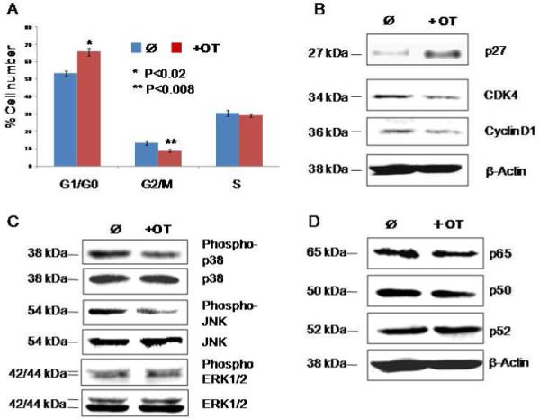
Figure 1.

OT causes cell cycle arrest in MIA cells through the MAPK pathway. Panel A shows the alterations of cell cycle distribution by OT treatment as measured by flow cytometry. Panel B shows the altered activity of cell cycle regulators by OT treatment as measured by Western blot. Panel C shows the inhibition of phosphor-p38 and JNK in the MAPK cascades by OT treatment as measured by Western blot. Panel D shows the effect of OT treatment on NF-κB pathway as measured by Western blot. Please see the Materials and Methods section for details. All the experiments were repeated three times.
