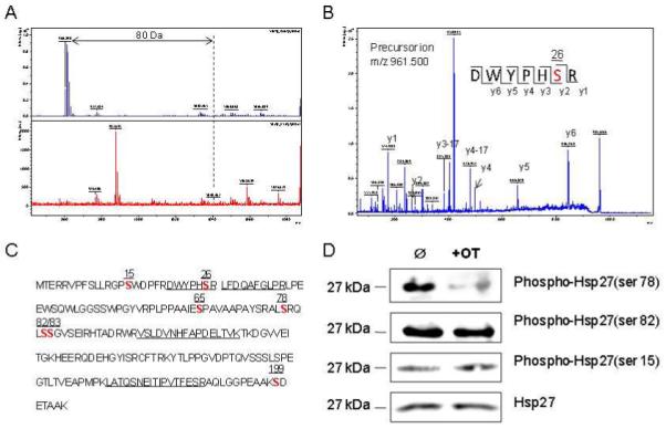
Figure 4.

Identification of the phosphorylation site of Hsp27. Panel A shows determination of the phosphorylated site of the peptide by de-phosphorylation with CIAP (Calf Intestinal Alkaline Phosphatase, upper panel in A) and without CIAP (lower panel in A). Panel B shows the MS/MS spectrum of this phosphor fragment in ‘lift” mode. The peptide sequence is easily identified as almost every y ion is observed. Panel C shows the whole sequence of human Hsp27. The number on the top of each bold “S” indicates the possible phosphorylated serine in this protein. Panel D shows overexpression of phosphor Hsp27 at Ser78, Ser82 and Ser15 in MIA cells treated with and without OT by using Western analysis. Please see the Materials and Methods section for details.
