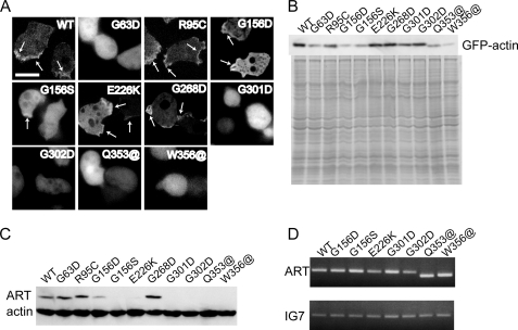
FIGURE 2.
Expression of GFP-mutant actins and mutant ARTs in Dictyostelium cells. A, fluorescence from GFP-actins in Dictyostelium cells observed using a confocal laser-scanning microscope. For GFP-G156D, -G156S, -Q353@, and -W356@ mutant actins, the majority of cells were too dark for observation, and the cells shown here have atypically high expression. Regions of cortical GFP fluorescence are indicated by arrows. Scale bar, 10 μm. B, Western blot (top) and Coomassie Brilliant Blue staining (bottom) following SDS-PAGE of total cell lysates from Dictyostelium cells expressing GFP-mutant actins. Polyclonal anti-GFP antibodies were used for Western blotting. C and D, levels of ART protein and mRNA were examined by Western blot analysis using a monoclonal anti-actin antibody (C) and RT-PCR using ART-specific primers (D).

