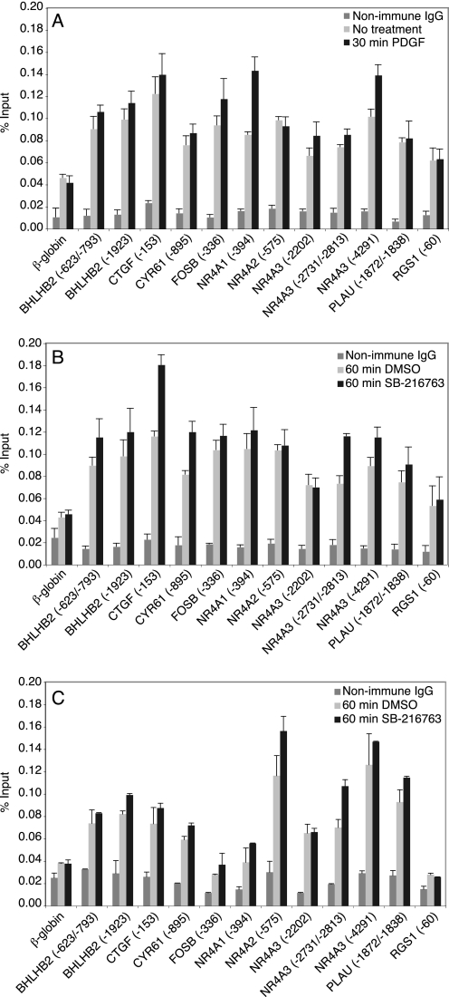
FIGURE 3.
Binding of p50 and HDAC-1 to predicted NF-κB sites in quiescent and stimulated cells. Quiescent T98G cells were either untreated or treated for 30 min with PDGF (A) or treated for 60 min with either DMSO or SB-216763 (B and C). Chromatin fragments were precipitated with either anti-p50 antibody (A and B), anti-HDAC-1 antibody (C) or nonimmune IgG and quantified by real time PCR. The values for nonimmune IgG are from treated cells. The data are presented as the percentage of input and are the means of two (C) or three (A and B) independent experiments ± S.E.

