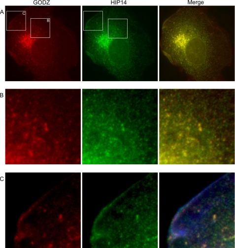
FIGURE 3.
Subcellular localization of GODZ and HIP14 proteins. Shown is the immunofluorescence staining of GODZ-FLAG and HIP14-GFP fusion proteins in transiently expressing COS-7 cells. A, Golgi localization of GODZ. The merged image is shown. B, presence of GODZ-FLAG and HIP14-GFP in Golgi and post-Golgi vesicles. Images are 4× magnifications of the boxed areas in A. C, subplasma membrane location of post-Golgi GODZ-FLAG and HIP14-GFP in cells stained with phalloidin to mark the plasma membrane ruffles. Images are 4× magnifications of the boxed areas in A. Note the evident subplasma membrane localization of both GODZ and HIP14 proteins in this region of normal cells.

