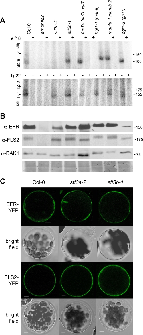
FIGURE 2.
Ligand binding, protein accumulation, and localization of PRRs in selected N-glycosylation mutants. A, equal amounts of ground seedling material were incubated with elf26-Tyr-125I or 125I-Tyr-flg22 in the absence (−) or presence (+) of 10 μm elf18 or flg22 peptide, respectively. Similar results were obtained in at least three independent experiments. B, accumulation of EFR, FLS2, and BAK1 proteins. Equal amounts of the lines indicated in A were loaded for immunoblotting and revealed with specific antibodies (α-FLS2, α-EFR, or α-BAK1). As controls, efr mutants were included for elf26 binding and EFR and BAK1 accumulation, and fls2 mutants were included for flg22 binding and FLS2 accumulation. Representative Coomassie staining is shown as loading reference below. C, EFR-YFP and FLS2-YFP were expressed in protoplasts of Col-0 wild type and indicated glycosylation mutants. Confocal and bright field images of representative transfected protoplasts are shown. Scale bars, 5 μm.

