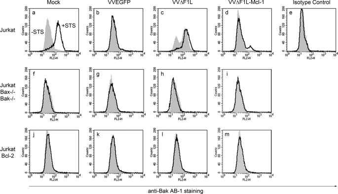
FIGURE 9.
Mcl-1 inhibits Bak activation during VVΔF1L infection. Jurkat cells (a–e), Jurkat cells devoid of Bax and Bak (f–i), and Jurkat cells overexpressing Bcl-2 (j–m) were mock-infected or infected with VVEGFP, VVΔF1L, or VVΔF1L-Mcl-1 for 4 h at an m.o.i. of 10 before treatment with 250 nm STS for 1.5 h to induce apoptosis. Bak N-terminal exposure was monitored by staining cells with the conformation-specific anti-Bak AB-1 antibody or anti-NK1.1 as an isotype control antibody. Shaded histograms, untreated cells; open histograms, STS-treated cells.

