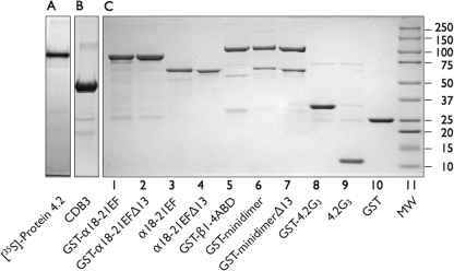
FIGURE 1.
Proteins used in this study separated on 9% Laemmli-Tricine SDS gels (7). A, shown is an autoradiograph of the in vitro transcription/translation product of full-length (77 kDa) protein 4.2, labeled with [35S]methionine. B, shown is the cytoplasmic domain of band 3 cleaved and purified from red cell membranes stripped of peripheral proteins (17). C, shown are recombinant proteins used in this study. 8 μg of each protein were analyzed. In some cases (lanes 3, 4, and 9) the GST moiety had been cleaved from the recombinant fusion proteins with PreScission protease (Amersham Biosciences) before electrophoresis.

