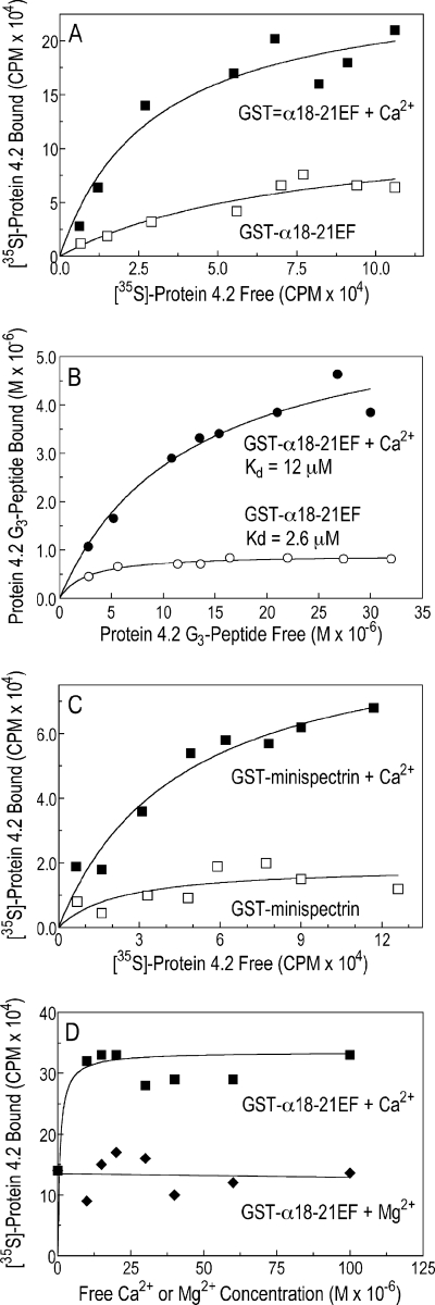
FIGURE 8.
Binding of protein 4.2 to the α-spectrin EF-domain is amplified by Ca2+ but not by Mg2+. A, shown is binding of 35S-labeled protein 4.2 to GST-α18–21EF in the absence or presence of free Ca2+ (1.5 mm). B, shown is binding of the 125I-labeled G3 peptide of protein 4.2 to GST-α18–21EF (0.75 μm) in the absence or presence of free Ca2+ (1.5 mm). C, shown is binding of 35S-labeled protein 4.2 to GST-minispectrin (0.43 μm) in the absence or presence of free Ca2+ (1.5 mm). D, shown is the specificity of Ca2+ compared with Mg2+ in the binding of 35S-labeled protein 4.2 to GST-α18–21EF (0.75 μm). All experiments used the HEPES binding buffer, which contains 0.5 mm EGTA (see “Experimental Procedures”) with or without added CaCl2 or MgCl2. Free Ca2+ and Mg2+ concentrations were calculated using the EGTA Calculator.

