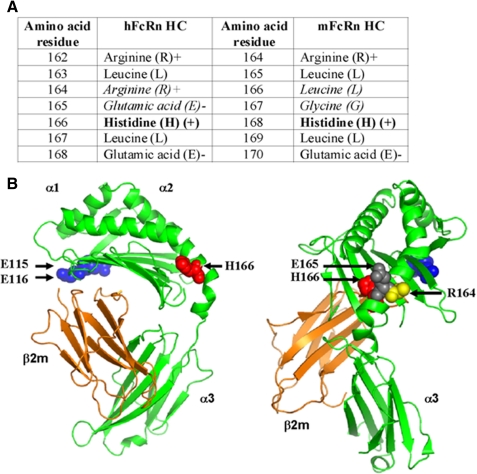
FIGURE 7.
The crystal structure of shFcRn. A, amino acids flanking His-166 (hFcRn) and His-168 (mFcRn) located within the heavy chain α2-domain are shown. His-166 and His-168 are shown in bold. The non-conserved Arg-164 and Glu-165 of hFcRn, and Leu-167 and Gly-168 of mFcRn are shown in italic. B, the crystal structure of shFcRn shown in two orientations. The localization of amino acids essential for IgG (Glu-115 and Glu-116) and albumin (His-166) binding are highlighted as blue and red spherical balls. The non-conserved Arg-164 and Glu-165 (human) are highlighted with yellow and gray spherical balls, respectively. The FcRn heavy chains are shown in green and the β2m in orange. The figures were designed using PyMOL (DeLano Scientific) with the crystallographic data of shFcRn (37).

