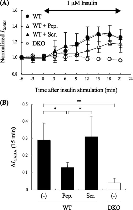
FIGURE 5.
Effect of PRIP1-(553–565) peptide on insulin potentiation of IGABA in hippocampal CA1 neurons. A, PRIP1-(553–565) peptide (3 μg/ml) (open triangles, n = 3), which diminishes the binding between PRIP and the GABAA receptor β-subunit (29), or its scramble peptide (3 μg/ml) (closed triangles, n = 3) were introduced using a patch pipette. The experiment was performed in the same way as that shown in Fig. 1A. A double-headed arrow indicates the time period of insulin application. Data are represented by the means ± S.D.. The IGABA from WT (closed circles, dashed line) or DKO (open circles, dashed line) mice without the peptide, which were taken from those shown in Fig. 1A, are also shown as references. B, graph shows the potentiation of IGABA at 15 min after insulin stimulation in WT (filled columns) or DKO (open column) neurons with or without the indicated peptides (Pep., PRIP1-(553–565 peptides); Scr., PRIP1-(553–565) scramble peptides; (−), no peptides). Data are represented as means ± S.D. Significance was determined using the Student's t test (*, p < 0.05; **, p < 0.01, between indicated two columns).

