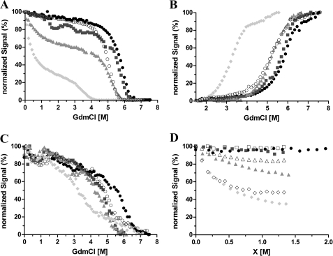
FIGURE 2.
Chemical denaturation of Venus monitored by the yellow fluorescence (A), tyrosine fluorescence (B), far-UV CD (C), and yellow fluorescence (D) of Venus as a function of GdmCl, urea, and NaCl concentration. A–C, 10 μm protein in 50 mm MES buffer at pH 6.0 (filled circles) and pH 6.6 (squares) or 25 mm Tris buffer for pH 7.6 (triangles) and pH 8.0 (diamonds). Measurements were taken after 7 days of incubation at 25 and 37 °C (open circles). The data are normalized relative to their individual maximum fluorescence intensity. D, normalized chromophore fluorescence intensity at 527 nm is plotted as a function of denaturant (closed symbols) or chloride ion concentration (open symbols). Aliquots of 10 μm Venus were incubated with various GdmCl or urea concentrations or NaCl in the absence of denaturants for 7 days prior to the measurements. The samples were in 50 mm MES buffer for pH 6.0 and pH 6.6 or 25 mm Tris buffer for pH 7.6.

