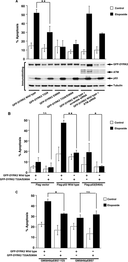
FIGURE 4.
ATM regulates DYRK2-mediated induction of apoptosis in response to DNA damage. A, U2OS cells were transfected with GFP-DYRK2 or its mutants and then treated with etoposide for 24 h. The percentages of apoptotic cells were quantified by TUNEL assays. The results are represented as the percentage of TUNEL-positive cells out of a total of GFP-positive cells. Values indicate the mean ± S.D. from three independent experiments. Cells were also analyzed by immunoblotting with indicated antibodies. Statistical analysis was performed with Student's t test. Double asterisks indicate p < 0.01. B, HCT116/p53−/− cells were transfected with FLAG vector, FLAG-p53, or the FLAG-p53 S46A mutant and GFP-DYRK2 or the GFP-DYRK2 mutant and then treated with etoposide for 24 h. Apoptotic cells were analyzed by TUNEL assays as described above. Double asterisks, the single asterisk, and n.s. indicate p < 0.01, p < 0.05, and not significant, respectively. C, GM 5849/pEBS7 or GM 5849/pEBS7-YZ5 cells were transfected with GFP-DYRK2 wild type or the T33A/S369A mutant followed by treatment with 20 μm etoposide for 24 h. The percentages of apoptotic cells were quantified by TUNEL assays. The results are represented as the percentage of TUNEL-positive cells out of a total of GFP-positive cells. Values indicate the mean ± S.D. from three independent experiments. The single asterisk and n.s. indicate p < 0.05 and not significant, respectively.

