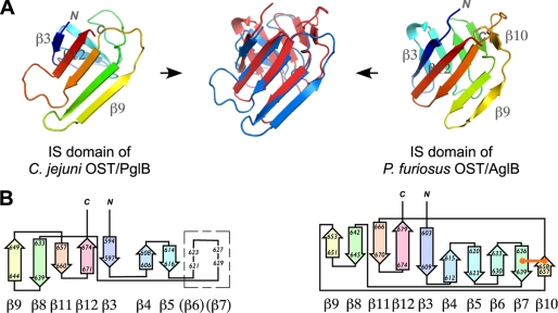
FIGURE 3.
Structural comparison of the IS domains of PglB and AglB. A, the overlaid structures (blue, PglB; red, AglB) are placed in between the IS structures of PglB (left) and AglB (right). B, topology diagram of the IS domains, generated by the PDBsum server. The location of the two putative β-strands suggested by structural similarity between PglB and AglB in a region with high temperature factors is shown in a box. The disulfide bond is shown as an orange line.

