FIGURE 1.
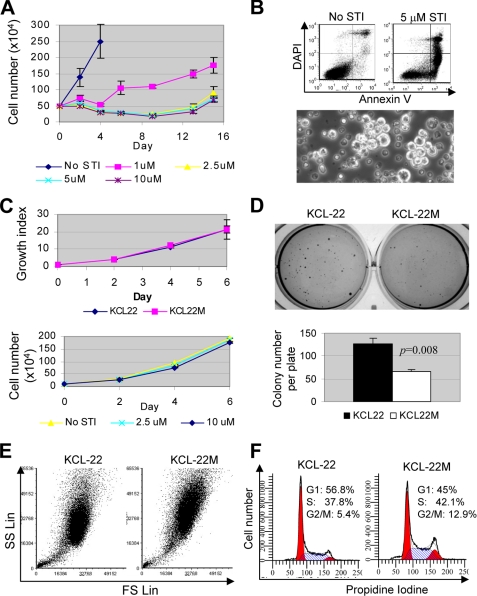
Novel model of CML acquired resistance. A, one-half million KCL-22 cells were treated with 1, 2.5, 5, and 10 μm imatinib (STI), and at days after the treatment as indicated, survival cells were counted. Relapse occurred on 2.5 μm and higher concentrations of imatinib 2 weeks post-treatment. B, top, apoptosis of KCL-22 cells after 6 days of treatment with 5 μm imatinib was analyzed by annexin V staining. DAPI, 4′,6-diamidino-2-phenylindole. Bottom, formation of clusters of resistant cells (bright) among scattered dead cells (dark) with 2.5 μm STI-571 treatment for 8 days. C, top, growth curves for resistant cells (KCL-22M) and KCL-22 cells analyzed by XTT. Growth indexes were relative XTT readings normalized to the initial XTT readings at day 0. Bottom, comparison of growth of KCL-22M cells in the absence and presence of imatinib. D, soft agar colony formation of KCL-22 and KCL-22M cells. Five hundred cells were seeded each well in 6-well plates in triplicate. E, comparison of cell size and complexity of KCL-22 and KCL-22M cells. KCL-22M cells exhibited increase at both forward scatter (FS) and side scatter (SC) parameters. SS Lin, side scatter in linear scale. F, comparison of cell cycle of KCL-22 and KCL-22M cells with propidium iodine staining.

