Fig. 3.
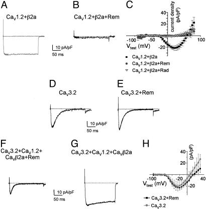
Rem and Rad prevent de novo expression of CaV1.2 ionic current but not T type Ca2+ currents. (A) Representative Ba2+ current elicited by a voltage step from –80 to 10 mV in HEK293 cells transiently transfected with expression vectors encoding CaV1.2, β2a, and GFP. (Bars = 10 pA/pF and 50 ms.) (B) HEK293 cells cotransfected with CaV1.2, β2a, and GFP-tagged wild-type Rem or Rad (same conditions as A). No current is detectable. (C) Current–voltage relationships for cells transfected with CaV1.2, β2a, and unfused GFP (filled squares) or CaV1.2, β2a, and GFP-Rem (filled circles) or Rad (open triangles). Average and SE from six to eight experiments for each group are shown. (D and E) Representative control CaV3.2 ionic current and CaV3.2 ionic current coexpressed with GFP-Rem. Also, coexpression of β2a with CaV3.1 or 3.2 or coexpression of CaVβ2a, GFP-Rem, and CaV3.1 or 3.2 had no effect on T type Ca2+ channel current expression (data not shown for CaV3.1). (F and G) Coexpression of CaV3.2, CaV1.2, β2a display a complex kinetic pattern that resolves to an ionic current that on Rem expression is indistinguishable from the CaV3.2 expressed alone. (H) The current–voltage curve shows that CaV3.2 current amplitude expression is not altered by Rem coexpression. Average and SE from six to eight experiments for each group are shown.

