
Figure 3.
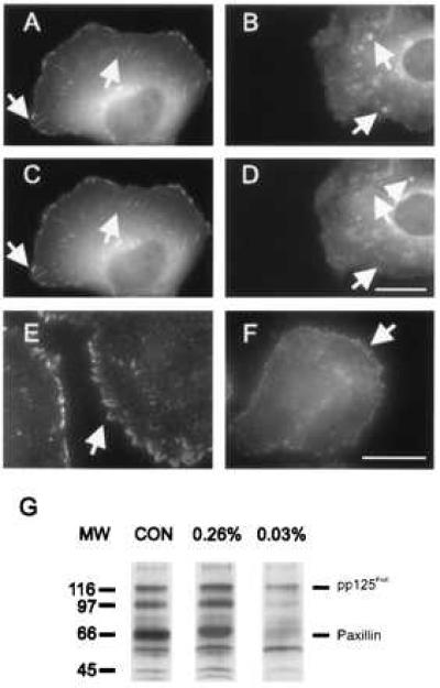
Distribution of vinculin and phosphotyrosine in NRK cells cultured on substrates with 0.26% bis-acrylamide (A, C, E) or 0.03% bis-acrylamide (B, D, F). (A–D) Cells were injected with rhodamine-labeled vinculin and imaged over a period of 10 min. On more rigid substrates (A, C), vinculin is incorporated into elongated focal adhesions, which show only minor changes during the period of observation. On highly flexible gels (B, D), vinculin is localized at punctate structures of irregular sizes and shapes, many of which appear and disappear over a period of 10 min (arrows). (E, F), Immunofluorescence of phosphotyrosine. Phosphotyrosine is localized at elongated focal adhesions in cells cultured on more rigid gels (E), and at punctate structures in cells cultured on highly flexible gels (F). (G) Anti-phosphotyrosine immunoblotting of whole cell lysates from NRK cells cultured on different substrates. On plastic dishes (CON) and rigid 0.26% bis-acrylamide substrates (0.26%), pp125FAK, paxillin and a 97-kDa protein are heavily phosphorylated after 48 hr of culture. Cells cultured on soft 0.03% bis-acrylamide substrates (0.03%) show a significantly lower extent of phosphorylation at these bands. Bar = 10 μm.