Figure 3.
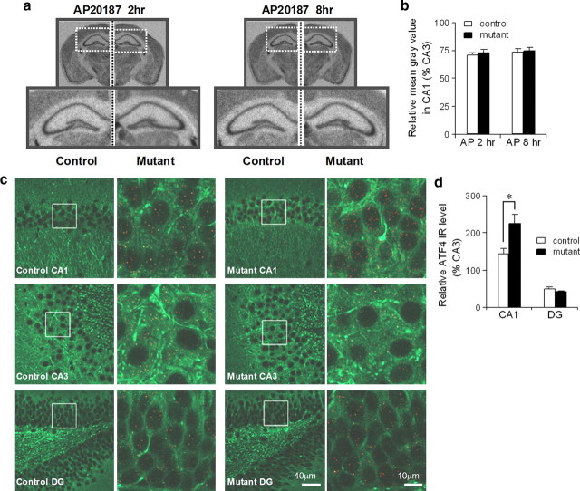
CA1-specific-increase in ATF4 expression without general translation inhibition upon elevation of eIF2α phosphorylation. a, b, No general translation inhibition was detected upon activation of PKR induced by AP20187 infusion. [35S]-methionine was intraperitoneally injected into mutants and fPKR controls 2 or 8 h after AP20187 infusion. The images below are enlargements of the boxed area in the upper images. De novo protein synthesis was quantified in NIH ImageJ by measuring the mean gray value in hippocampal CA1 and normalized to the level of CA3. c, d, Increased ATF4 expression was observed in mutant CA1 area but not in dentate gyrus (DG). Sections from fPKR control and mutant animals were double-immunostained with anti-ATF4 (red signal) and anti-MAP2 (green signal) and were image-collected by confocal microscope. The boxed areas from the left panel are enlarged in the right panel. The numbers of red dots for ATF4 signal in somatic area of CA1, dentate granule cells, and CA3 were counted using NIH ImageJ. The relative ATF4 values were normalized to the level of CA3 cell layer-IR. Mann–Whitney U test, *p < 0.05. Error bars represent SEM.

