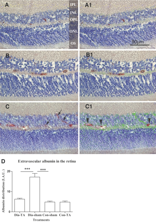
FIG. 4.
Immunohistochemical analysis of retinal albumin distribution. Albumin was detected (in red) within the vessels in sham-treated nondiabetic retinas (A). A similar pattern of albumin distribution was observed in IVTA-treated diabetic retina, but with slightly higher expression in extra vascular space (B). Diffuse extravascular albumin was observed in the inner nuclear, outer plexiform, and outer nuclear layers in sham-treated diabetic retina (C). Extravasated albumin (arrows in C) was marked green in panel A1, B1, and C1 (corresponding to panels A, B, and C) by IPP4.5. Intravascular albumin was excluded by the analysis (arrow heads in C1). D: Quantification of albumin distribution/leakage in the four treatment groups. Values were presented as IAU. Data are means ± SEM, n = 8; *** P < 0.001. Original magnifications ×400. To view a high-quality digital representation of this image, go to http://dx.doi.org/db07-0982.

