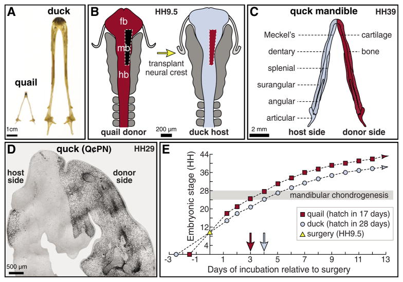
Fig. 1. Experimental design and methods.
(A) Lower jaw skeletons of adult Japanese quail (Coturnix coturnix japonica) and white Pekin duck (Anas platyrhyncos). (B) Schematic of rostral neural tube at HH9.5, depicting the levels of neural crest cells grafted from quail to duck. (C) Schematic of a lower jaw skeleton at HH39, depicting the contributions of transplanted neural crest (red) to cartilage and bone (stippled). (D) Horizontal section through the mandibular primordium of a HH29 chimeric quck embryo (rostral at top), which will give rise to the lower jaw skeleton. Quail donor mesenchyme (black), as visualized by the quail-specific antibody Q¢PN, was found throughout the transplanted side, whereas few to no quail cells were observed on the contralateral duck host side. (E) Graph illustrating the distinct developmental trajectories of quail (red squares) versus duck (blue circles) after being stage-matched at HH9.5 for surgery (yellow triangle on y-axis). Control quail and duck embryos were separated by approximately three HH stages within 2 days of surgery, and throughout the initial stages of overt mandibular chondrogenesis (gray area).

