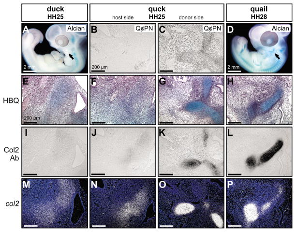
Fig. 4. Mesenchyme regulates late histogenesis of Meckel’s cartilage.
(A) Whole-mount Alcian Blue stained embryos at HH25 reveal that cartilage has yet to form in proximo-lateral regions of the avian mandible (arrow). (B) Duck host mesenchyme was negative for the anti-quail antibody Q¢PN, as shown in sagittal section. (C) By contrast, donor sides of HH25 chimeric quck mandibles contained abundant quail neural crest-derived mesenchyme. (D) Jaw cartilages became obvious by HH28 (arrow). (E) HBQ-stained histological sections through the jaw joint of control embryos at HH25 revealed diffuse Alcian Blue staining in mesenchyme with ill-defined borders. (F) Similar low diffuse levels of Alcian Blue were observed in host sides of HH25 quck mandibles. (G) In conjunction with the presence of relatively older quail donor cells, developing cartilages of quck chimeras stained strongly with Alcian Blue and exhibited a defined perichondrium. (H) Robust Alcian Blue staining and a clear perichondrium characterized developing cartilages at HH28. (I) Mesenchyme of the mandible was not immunoreactive for Collagen type II protein (Col2) in control embryos at HH25. (J) The host sides of chimeras were also negative for Col2 protein. (K) Quail-derived mesenchyme of HH25 quck mandibles demonstrated strong Col2-immunoreactivity. (L) Control HH28 mandibular cartilages were also positive for Col2 protein. (M) col2a1 expression appeared diffuse in developing mandibular cartilages in control HH25 embryos. (N) The host side of quck at HH25 also showed low levels of col2a1 expression. (O) Quail-derived mesenchyme of HH25 chimeric mandibles had more spatially resolved col2a1 domains, as well as higher col2a1 expression levels, when compared with contralateral duck host mesenchyme. (P) Similar expression domains were observed at HH28 in control quail.

