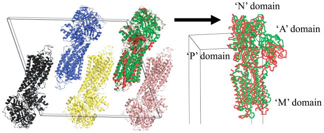
Fig. 7.
The comparison between crystal packing of the target conformation (1T5S) and the position and orientation of the final candidate conformation morphed from 1SU4 after applying MR. Though the N, P, and M domains are determined properly, however, the domain A has positional error, while the orientation of the domain is placed near correct orientation. The target conformation is presented as green color, the final candidate conformation as red.

