
Figure 10.
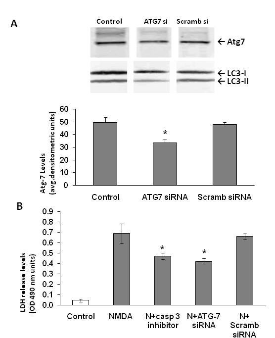
Knockdown of ATG7 results in neuroprotection following NMDA-exposure. A) ATG7 siRNA was transfected into neurons 72 hours before NMDA treatment. Representative western blot demonstrates the knockdown of the ATG7 in neurons (ATG7 siRNA) compared to controls and scrambled siRNA (Scramb siRNA). Representative LC3 immunoblot also show reduction of LC3-II band. Quantification of the band intensities (n = 3) representing the Atg7 protein levels in the granule neurons demonstrates a significant reduction in Atg 7 protein expression. Scrambled siRNA was used as a negative control to compare the efficiency of Atg7 protein suppression in neurons. *p < 0.05 ANOVA, Atg7 protein levels are significantly lower in the siRNA paradigm compared to controls and scrambled siRNA B) Lactate dehydrogenase (LDH) release from the neurons into the medium was measured and quantified at 6 hours post treatment (n = 3). *p < 0.05, ANOVA, NMDA+caspase-3 inhibitor (IDN- and NMDA+ATG7 siRNA treatment groups are significantly lower compared to NMDA treated neurons. Data is represented as mean ± S.E.M.