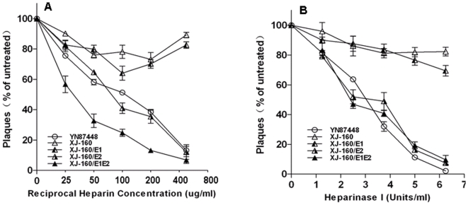
Figure 4. Effect of heparin or heparinase I treatment on plaque formation by the chimeric or parental viruses.
(A) Viruses diluted to 100–200 PFU/200 µl were incubated with heparin at the concentrations indicated for 1 h at 37°C. Then plaque assays were performed using BHK-21 cells as described in the Materials and Methods. (B) Confluent BHK-21 cell monolayers were treated with heparinase I at the concentrations indicated. After washing three times with PBS, the cells were infected with viruses diluted to 100–200 PFU in 200 µl. Plaque formation was analyzed as described.

