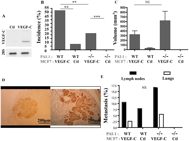
Figure 1. Tumor development after orthotopic injection of VEGF-C overexpressing MCF7 cells or control MCF7 cells implanted in the mammary fat pads (mfp) of PAI-1 WT or PAI-1−/− mice.
(A): RT-PCR analysis of VEGF-C and 28S mRNA expression by MCF7. (B): Tumor incidence (%) is defined as the percentage of palpable tumor per mfp. (C): Tumor volume was measured as described in Material and Methods. (D): Representative figure of a typical metastasis in lymph node (left) and lung (right). (E): Percentage of animal bearing at least a tumor nodule (metastasis% detected in lymph nodes (black boxes) and lungs (white boxes). Number of mfp per condition = 34–38. The mice PAI-1 status (WT or −/−) and the VEGF-C production (VEGF-C) or not (Ctl) by MCF7 cells are indicated below each graph. Data are ± S.E.M. Scale bars: 200 µm. ** P≤0.01, *** P≤0.001, NS = Non Significant.

