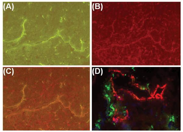
FIGURE 5. Expression of MHC antigens on lymphatic endothelium. DA-to-Lewis allograft, post-transplant day 7.
(A) Lymphatic vessels labeled with green immunofluorescence (LYVE-1 and Alexa-488). (B) Lymphatic vessels in (A) doubly labeled with red immunofluorescence for MN4-91-6, a polymorphic determinant of MHC I. (C) Overlay of (A) and (B) demonstrated co-expression of LYVE-1 and MHC I on the lymphatic endothelium. (D) Immunofluorescent label of MHC II with OX6 (green), a monomorphic determinant of rat MHC class II, showed that MHC II expression did not overlap with LYVE-1-positive lymphatic endothelial cells (red). Magnifications: X200 in A through C. X400 in D.

