
Fig. 2.
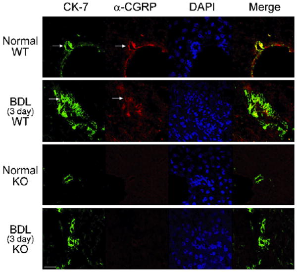
Localisation of α-CGRP (red) by immunofluorescence in normal and 3-day BDL WT mice liver sections. Bile ducts were stained with CK-7 (green). α-CGRP-positive staining and CK-7 colocalise in the bile ducts of 3-day BDL WT mice. The scale bar represents 20 μM. Arrows indicate bile ducts.
Reproduced with permission from Ref. [53].