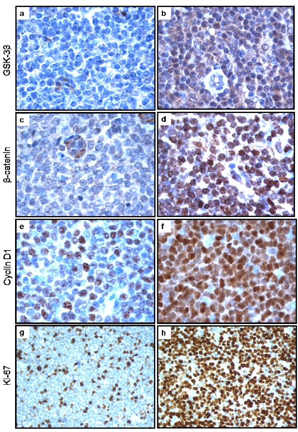
Figure 1.
a, b: Immunohistochemical staining of pGSK-3β in MCL tumors revealed a subset of negative cases and a subset of positive case (a and b respectively). The staining was predominantly cytoplasmic. c, d: Immunohistochemical staining of β-catenin in MCL tumors revealed a subset of negative cases and a subset of positive case (illustrated in c and d respectively). Of note, the staining in the tumor cells was predominantly nuclear; in contrast, the staining was mostly found in the cytoplasm of the endotheiiai cells. e, f: Immunohistochemical staining of cyclin D1 in MCL tumors was heterogeneous with regard to the proportion of intensely positive cells. The case in 1e was assessed cyclin D1-low, as strongly positive cells were <50% of the neoplastic cell population. The case in figure 1f was assessed cyclin D1-high, as strongly positive cells were ≥ 50% of the neoplastic cell population, g, h: Immunohistochemical staining of Ki67 in MCL tumors also revealed a high degree of heterogeneity in the proportion of positive cells, showing a Ki67-negative case and a Ki67 positive case (gand h respectively).

