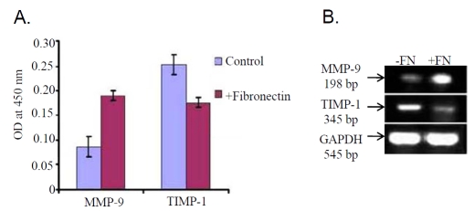
Figure 4.
(A) SFCM collected from K562 cells cultured in presence (+Fibronectin) and in absence (Control) of 20μg/ml fibronectin was coated in microtitre wells. Wells were blocked with 1% BSA followed by incubation with anti-MMP-9 and anti-TIMP-1 primary antibody and respective secondary antibodies. OD was taken at 450nm as described above. (B) Total RNA was extracted from 20μg/ml fibronectin treated (lane +FN) and untreated (lane -FN) K562 (300,000 cells/ml) cells. RT-PCR was done as described above using MMP-9 and TIMP-1 primer and bands were visualized in 2% agarose gel under UV. GAPDH was used to confirm total RNA integrity and equal loading.

