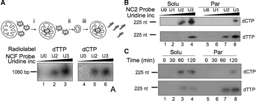
Figure 4.
DNA repair mechanisms differ dependent on their mitochondrial location. (A) Mitochondria repair uracil-containing DNA on import. In these import and repair experiments the isolated mitochondria were first (i) incubated with the free radionucleotide (dTTP lanes 1–3; dCTP lanes 4–6), (ii) harvested and (iii) washed twice to remove unincorporated radiolabel. Aliquots of 1060 bp probe were prepared with increasing levels of uracil incorporation (U0–U3) as detailed in ‘Materials and Methods’ section and incubated with the pre-loaded mitochondria in DNA repair buffer (RB) for 45 min before DNase I treatment, DNA extraction, separation through non-denaturing gels, transfer to nylon membranes and visualization. (B) Mitochondrial sub-fractions show differing DNA repair mechanisms. Aliquots of probe NC2 were prepared with increasing amounts of uridine prior to incubation with pre-loaded mitochondria in RB for 2 h. Following import, mitochondria were sub-fractionated and incorporation of radiolabel into the probe in each fraction was monitored after migration under denaturing conditions and autoradiography. Upper panel, mitochondria were pre-loaded with dCTP; lower panel, dTTP. (C) Repair driven DNA synthesis as a function of time. A similar experiment was performed with pre-loaded mitochondria incubated for the indicated times with probe NC2U3. Mitochondria were sub-fractionated, DNA isolated and analysed exactly as described in B.

