
Figure 4.
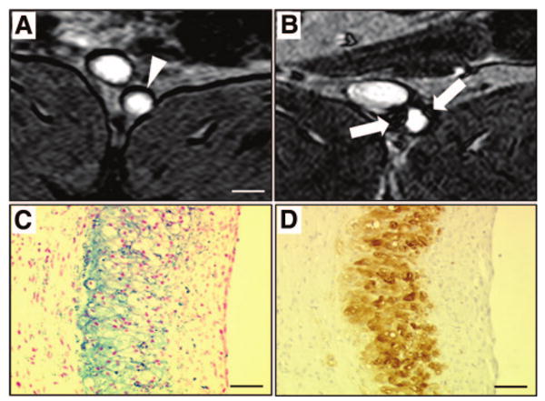
Detection of macrophages with USPIO-enhanced MRI. MR axial views of an atherosclerotic plaque in a rabbit aorta before (A) and 5 days (B) after the intravenous injection of ultrasmall superparamagnetic iron oxide nanoparticles (USPIO). Five days after the intravenous injection of USPIO, strong signal voids (white arrows) were detected in the aortic wall using T2*-weighted MR sequences. Note the dark artifacts that are not related to USPIO accumulation at the fat-water interfaces. On corresponding histological section, accumulation of iron oxide nanoparticles was identified in the atherosclerotic plaque with Perls stain (C; blue staining for iron) and colocalized with macrophage infiltration detected by immunohistotochemistry using a RAM-11 monoclonal antibody for rabbit macrophages (D; brown staining). White scale bar, 5 mm; black scale bar, 30 μm.