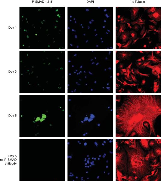
Figure 5. Phosphorylated SMAD1,5,8 Immunofluorescence.
Osteoclasts were differentiated by RANKL for 1, 3 or 5 days and subjected to immunofluorescence staining against phosphorylated SMAD1,5,8 (green). Cells were costained against α-tubulin (red) and with DAPI (dark blue) to show cell outline and nuclei, respectively. Parallel slides from each day were stained without P-SMAD antibody as a negative control (bottom row and not shown). All images are at equal magnification and were acquired and processed identically.

