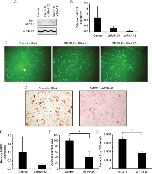
Figure 7. Suppression of BMPR II expression inhibits osteoclast formation.
A–B. Western blot (A) and real-time RT-PCR (B) showing expression of Type II BMP Receptor in RAW264.7 cells following infection 20ith control shRNA or BMPR II shRNA lentiviral vectors and puromycin selection. C. Fluorescence micrographs showing BMPR II and control-infected RAW264.7 cells that have been stimulated with RANKL for 6 days. Note the large multinucleated osteoclast indicated by the arrowhead in the “Control” panel. D. TRAP staining of Control and BMPR II shRNA -infected primary osteoclasts treated with RANKL for five days. E. Real time RT-PCR analysis of BMPR II expression in shRNA primary osteoclasts, plotted relative to control shRNA cells. F–G. Quantitative analysis showing the number (F) and size (G) of differentiated BMPR II suppressed primary osteoclasts following RANKL differentiation. *, p≤0.002.

