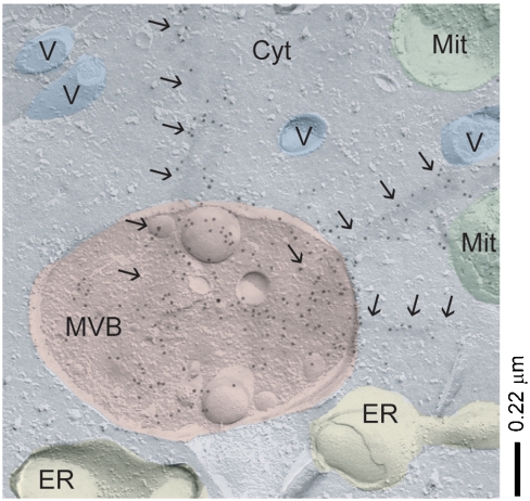
Fig. 5.
Aβ-dependent perturbation of the MVBs. TEM images of freeze-fractured J774A.1 cells showing Aβ-positive staining (immunogold labels) within MVBs and membrane penetration through fibrillar Aβ bundles (arrows). Possible assignments of other vesicular structures (V), mitochondria (Mit), endoplasmic reticulum (ER) and cytoplasm (Cyt) shown.

