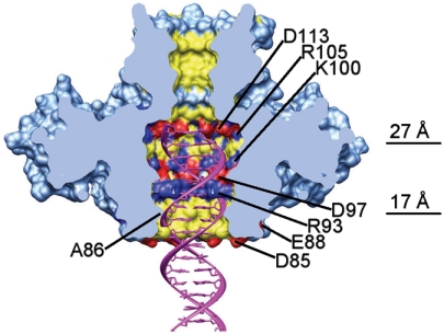
Fig. 4.
Structure of the gp1 channel. The molecular surface of the gp1 assembly is shown in light blue. The positive, negative, and hydrophobic residues in the channel are colored in blue, red, and yellow, respectively. Charged residues forming rings in the channel are labeled. The widest position of the channel in the gp1 body and the position of the constriction formed by residues A86 are indicated by lines and values of diameters. The front half of gp1 was computationally removed to show the internal surface of the channel. Also shown is a B-form double-stranded DNA (Magenta) fitted into the channel.

