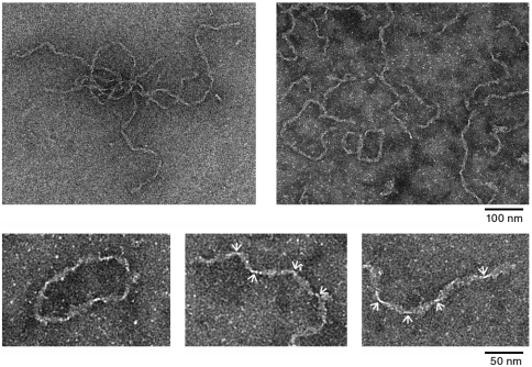
Fig. 4.
Transmission electron microscope images of MetO-apoA-I fibrils indicating the gross morphology of the fibrils. Small panels show magnified views of the fibrils including an example of the, apparently, circular fibrils. Constrictions in the fibril length due to the helical twisting of the fibril ribbon are indicated with arrows.

