
Fig. 2.

Comparison of the relative orientation of CheX and CheY3 with that of YPD1 (a histidyl phosphotransferase) and SLN1-R1 (a receiver domain) within  (pdb 2R25). For
 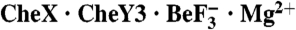 (Upper), CheX is gray except for α1′, which is cyan. CheY3 is green except α1 is blue and α5 is red. For
  (Lower), YPD1 is gray except for αC (teal). SLN1-R1 is pale yellow except α1 is blue and α5 is red.