Fig. 2.
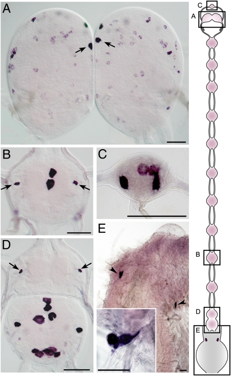
Expression of PTSP in the nervous system. (A–D) Brain with medial neurosecretory cells (arrows in A), fifth abdominal ganglion with interneuron 704 (IN704; arrows in B), frontal ganglion (C), and terminal ganglion with IN704 (arrows in D) of day 2 fifth-instar larva. (E) Epiproctodeal glands (arrowheads and Inset) of the fourth-instar larva (10 h before ecdysis). (Scale bar, 100 μm.) Schematic illustration of the nervous system is shown on the Right.

