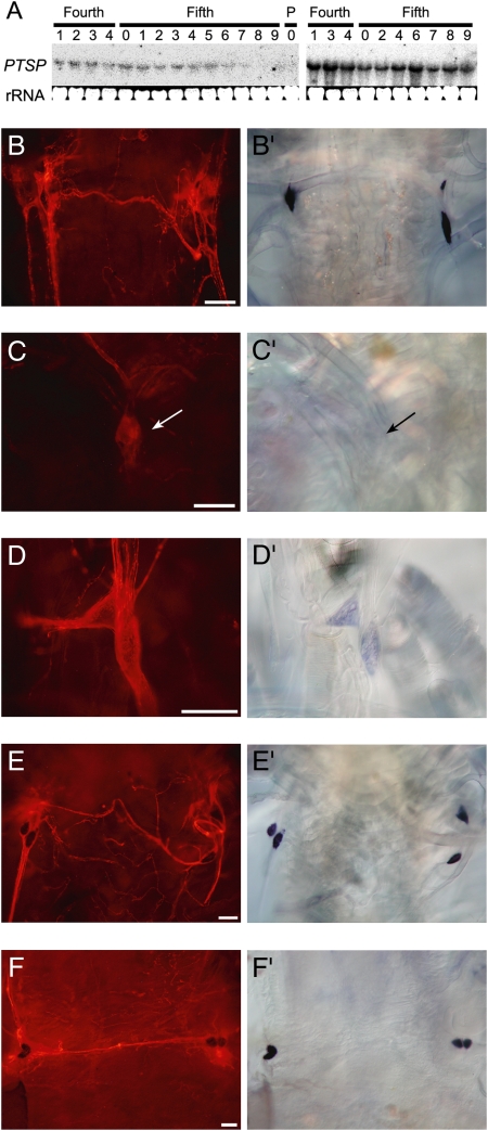
Fig. 3.
Temporal expression patterns of PTSP. (A) Northern blot analysis of PTSP expression during the fourth and fifth instars in the brain (Left) and the terminal abdominal ganglion (Right). (B–F) Immunostaining and (B′–F′) in situ hybridization (of the epiproctodeal glands. (B and B′) Fifth instar day 0 (just after ecdysis). (C and C′) Fifth instar day 1 (first day of feeding). (D and D′) Fifth instar day 6 (1 day before spinning). (E and E′) Fifth instar day 8 (1 day after the initiation of spinning). (F and F′) Fifth instar day 10 (just before pupal ecdysis). (Scale bar, 100 μm.)

