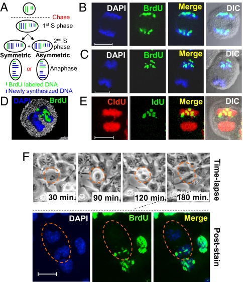
Fig. 1.
DNA is partitioned asymmetrically during mitosis in lung cancer cells. (A) Schematic representation of symmetric and asymmetric division of template DNA. Cells are grown in BrdU (green). During anaphase of the second cell division after BrdU is removed (the chase), the BrdU- labeled template chromosomes are segregated either randomly (symmetric) or exclusively to one side of the metaphase plate (asymmetric). (B–D) Representative anaphase A549 cells that partition their BrdU-labeled template DNA (green) either randomly to both top and bottom daughter cells (B) or exclusively to the top daughter cell (C and D). (E) An A549 cell asymmetrically partitions IdU-labeled template DNA (green) to the top daughter cell and randomly segregates the newly synthesized CldU-labeled DNA (red) to both daughter cells. (F) Time frames from live time-lapse imaging show a dividing label-retaining cell, outlined by a dotted circle. The two daughter cells, outlined by a dotted oval, were fixed and stained for BrdU. The BrdU-labeled template DNA was segregated exclusively to the upper daughter cell. DNA was stained with DAPI (blue). (Scale bar: 10 μm in B, C, E, and F.)

