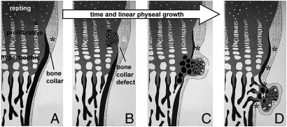
Fig. 6.
Osteochondromagenesis. (A) An early proliferative chondrocyte with homozygous disruption of Ext1 has no HS on its cell surface or in the immediate extracellular matrix (black clone of cells). (B) This loss of HS delays the clone’s differentiation and gives it a proliferative advantage. The immediately adjacent perichondrium fails to differentiate into osteoblasts and form a bone collar (white asterisk) due to blocked osteogenic signals from the physis, physical displacement away from the osteogenic signals from the physis, or receipt of alternate signals from the less-differentiated chondrocytes of the early osteochondroma. (C) Once the clone has progressed past the hypertrophic zone, the physis-flanking perichondrium receives normal osteogenic signals (black asterisks) again and reforms the bone collar in the wake of the osteochondroma. The osteochondroma may or may not have brought along cells of normal genotype (white cells, C and D) from the physis. (D) The fully developed osteochondroma, once completely independent from direct physeal signaling, begins to organize itself loosely into the proliferative (P) and hypertrophic (H) zones of a growth plate, as well as form its own primary spongiosa and bone collar, each in continuity with the host bone.

