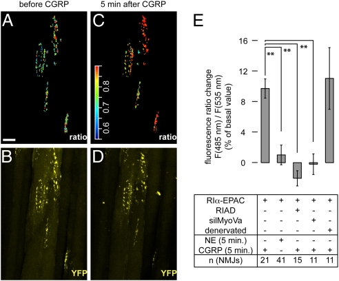
Fig. 3.
In the presence of exogenous CGRP, rise in subsynaptic [cAMP] is measurable upon denervation, but not upon impairment of AKAP or myosin Va function. TA muscles were transfected with RIα-EPAC or cotransfected with RIα-EPAC and RIAD (RIAD) or silMyoVa (silMyoVa). Some muscles were transfected with RIα-EPAC and denervated (denervated). Ten days later, muscles were injected with BGT-AF647 and monitored with in vivo two-photon or confocal microscopy. (A–D) Representative experiment with a RIα-EPAC-transfected muscle. Images depict 3D projections of the same NMJs before (A and B) and 5 min after injection of 50 μL of 10 μM CGRP (C and D). (A and C) Subsynaptic RIα-EPAC CFP/YFP ratio signals in pseudocolors. (Scale bar, 50 μm.) Color scale bar, pseudocolors corresponding to CFP/YFP ratio values. (B and D) YFP signals (yellow). (E) Quantification of different experiments. Shown is the percentage change in CFP/YFP ratio values [F(485nm)/F(535nm)] compared to basal values upon application of 50 μL of either 10 μM NE or 10 μM CGRP (indicated) ± SEM. Muscles were transfected or denervated as indicated. n-values indicated, ≥ three mice per condition.

