Fig. 4.
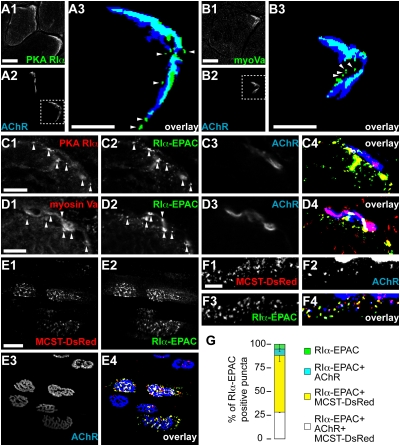
Myosin Va and PKA RIα share the same subsynaptic compartment. (A–D) TA muscles were snap-frozen, sectioned transversally, and immunostained against a C-terminal epitope of PKA RIα or myosin Va. AChRs were labeled with BGT-AF647. Confocal sections depict representative fluorescence signals as indicated. [Scale bars, 50 μm (A1 to A2, B1 to B2) or 10 μm (A3, B3, C, D).] (A3 and B3) Overlays, details showing the boxed regions in A2 and B2: immunostaining (green), AChR (blue). Fluorescence signals, binarized. (arrowheads) Punctate PKA RIα- or myosin Va-positive structures. (C and D) TA muscles were transfected with RIα-EPAC 10 days before staining. (arrowheads) RIα-EPAC-positive puncta double-positive for either PKA RIα (C) or myosin Va (D). Overlays, Fluorescence signals, binarized. PKA RIα/myosin Va (red), RIα-EPAC (green), AChR (blue). Double-positive structures: PKA RIα/myosin Va and RIα-EPAC (yellow); PKA RIα/myosin Va and AChR (magenta); triple-positive structures (white). (E–G) TA muscles were cotransfected with MCST-DsRed and RIα-EPAC. Ten days later they were injected with BGT-AF647 and monitored with confocal in vivo microscopy. (E1–E3) Representative 3D projections of fluorescence signals of markers as indicated. (Scale bar, 50 μm.) (F1–F3) Representative detail of a single optical section. Fluorescence signals of markers as indicated. (Scale bar, 10 μm.) (E4 and F4) Overlays of E1 to E3 and F1 to F3, respectively. Fluorescence signals, binarized. MCST-DsRed (red), RIα-EPAC (green), AChR (blue). MCST-DsRed colocalizing with RIα-EPAC (yellow). MCST-DsRed colocalizing with AChR (cyan). Triple-positive objects (white). (G) Quantification, fraction of RIα-EPAC-positive puncta also positive for AChR and MCST-DsRed (white), MCST-DsRed (yellow), AChR (cyan), or single positive (green). Mean ± SEM (n = 3 mice, 2,791 puncta analyzed).

