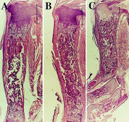
Figure 7.

Histological sections of decalcified tibiae from a newborn Tg-A/PTHrP+/+ mouse (A), a Tg-A/PTHrP−/− (B) littermate (both animals were homozygous for the transgene), and a representative newborn PTHrP−/− (C) mouse. The sections were stained with hematoxylin and eosin. The height of the proliferative chondrocyte layer is indicated by brackets. A–C show pictures at the same magnification.
