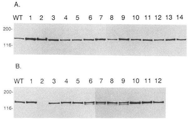
Fig. 2. Immunoblot and calmodulin overlay analyses of wild-type and mutant smooth muscle myosin light chain kinases.

Panel A, Western immunoblot analysis of full-length smooth muscle myosin light chain kinase measured in COS cell extracts by a monoclonal antibody directed against the bovine tracheal myosin light chain kinase. Approximately 20–40 ng of smooth muscle myosin light chain kinase was present in each lane. Molecular mass markers (kDa) are listed on the far left of the blot. Panel B, calmodulin overlay of smooth muscle myosin light chain kinases. Biotinylated calmodulin and horseradish peroxidase conjugated to avidin was used to detect smooth muscle myosin light chain kinases in COS cell extracts as described under “Materials and Methods.” Approximately 40 ng of smooth muscle myosin light chain kinase was loaded per lane. Molecular mass markers (kDa) are listed on the far left of the blots. For both panels A and B, WT is the wild-type, recombinant rabbit smooth muscle myosin light chain kinase; lanes 1–12 are K979E, RRK974–76EED, KK969–970EE, K965E-R967D, KK961–962EE, KK961–962EE/KK969–970EE, K965E-R967D/KK969–970EE, KK961–962EE/K965E-R967D, KK961–962EE/K965E-R967D/KK969–970EE, R967A, K965A, K965A-R967A, respectively. In panel A, lanes 13 and 14 are D966K and D966A, respectively. D966K and D966A also bind calmodulin using the identical overlay technique (data not shown).
