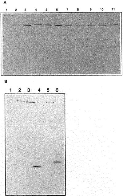
Fig. 4. Western immunoblot and calmodulin overlay analysis of chicken skeletal and smooth muscle MLCKs and mutant enzymes.
Purified MLCKs and COS cell extracts containing recombinant proteins were separated by 7.5% SDS-PAGE and detected with monoclonal antibodies or biotinylated calmodulin as described under “Materials and Methods.” Panel A, Western immunoblot with monoclonal antibody raised to chicken skeletal muscle MLCK. Lane 1, mock transfected COS cell lysate; lane 2, tissue-purified chickn skeletal muscle MLCK (10 ng); lanes 3–11, COS cell lysates transfected with 3, wild-type chicken skeletal muscle MLCK, 4, SK/SM1; 5, SK/SM4; 6, SK/SM6; 7, SK/SM8; 8, SK/SM2; 9, SK/SM9; 10, A494E 11, Δ515–516,K517E. Panel B, biotinylated calmodulin overlay. Lane 1, mock transfected COS cell lysate; lane 2, tissue-purified chicken skeletal myosin light chain kinase; lanes 3–6, COS cell lysates with 3, wild-type chicken skeletal muscle myosin light chain kinase; 4, SK-TR mutant; 5, SK/SM1 mutant; 6, SM-TR mutant.

