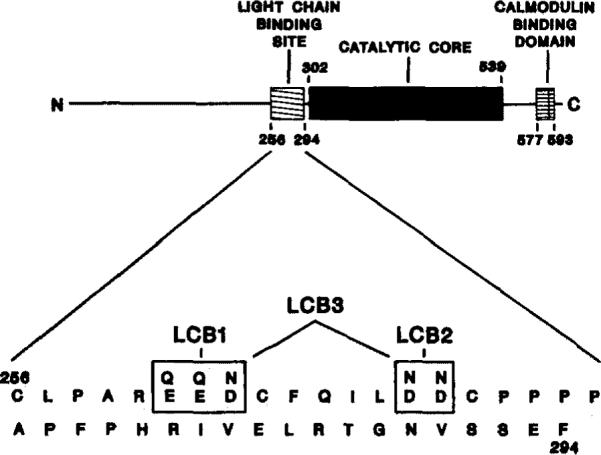
Fig. 1. Domain organization of and proposed light chain-binding site on rabbit skeletal muscle myosin light chain kinase.

A schematic representation of the linear amino acid sequence and domain organization of rabbit skeletal muscle myosin light chain kinase is shown (top). The catalytic core is the region homologous to other protein kinases (Hanks et al., 1988); the calmodulin-binding domain was defined by Blumenthal et al. (1985); a light chain-binding region is defined from results presented in this paper. The amino acid sequence of the proposed light chain-binding region is shown (bottom). The acidic residues (positions 261–263 and 269 and 270) which were altered in this present study are boxed. The substituted amino acids are indicated above the native residues. The nomenclature of the mutated kinases is also indicated.
