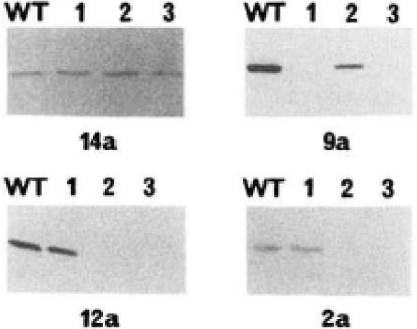
Fig. 3. Immunoblot analysis of mutant myosin light chain kinases.

Immunoblots were performed as described previously (Herring et al., 1990); ~20 ng of kinase were electrophoresed in each lane. The order of loading of each immunoblot was identical. Lane WT, wild-type kinase; lane 1, mutant LCB1; lane 2, mutant LCB2; lane 3, double-mutant LCB3. The immunoblots were reacted with the monoclonal antibody indicated below each blot.
