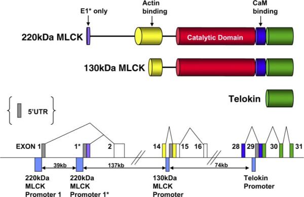
Fig. 1.
Structure of the mouse mylk1 gene. Top schematic is a representation of the domain structure of protein products of the mylk1 gene. As illustrated, the amino acid sequence of telokin is identical to the COOH terminus of the myosin light chain kinase (MLCK) molecules. Likewise, the amino acid sequence of the 130-kDa MLCK is identical to the common COOH-terminal portion of the 220-kDa MLCK. Bottom schematic is a representation of a portion of the mouse mylk1 gene (not to scale; see Supplemental Data for sequence information).2 Exons are indicated by boxes and are color coded to match the domains in the top schematic. The dark gray boxes represent the unique 5′-untranslated region (UTR) segments of each transcript. Promoter regions are indicated by the blue boxes below the line. Genomic sequence predicts that two 5′ promoters direct the expression of the two isoforms of the 220-kDa MLCK that differ from each other only in the sequence encoded by exon 1* of the 220-kDa MLCK E1*. The protein produced from usage of exon 1* is predicted to be 9 amino acids longer than the protein resulting from usage of exon 1. Two internal promoters direct the expression of transcripts encoding the 130-kDa MLCK and telokin. (For specific sequences, see Supplemental Data).

