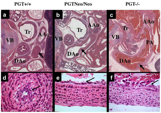
Figure 3. Patent DA in PGT Neo/Neo and PGT −/− mice.

H&E stain of paraffin-embedded sections.
(a) and (d). Low- and high-power view, respectively, of a cross-section from the torso of a PGT +/+ (wild type) mouse (representative, n = 3) eleven hours after birth. The DA has closed normally (a, arrow), and there is a normal intimal thickening (d, arrow) consisting of a loose network of cells filling and obliterating the constricted lumen. Tr, trachea; VB, vertebra; AAo, ascending aorta; DAo, descending aorta.
(b) and (e). Torso of PGT Neo/Neo mouse (representative, n = 5) dying on post-natal day 2 shows PDA. An arrow marks the connection between the DAo and DA. High power view (e) reveals normal intimal thickening (arrow).
(c) and (f). Torso of PGT −/− mouse (representative, n = 5) similarly shows patent DA. The pulmonary artery (PA) has dilated with reversed blood flow, and blood also fills the DAo and PA. High power (f) view reveals normal intimal thickening (arrow).
